Dinner Fork Deformity MEDizzy
If dorsal angulation is severe enough, a dinner fork deformity may be described.There is also usually impaction with resultant shortening of the radius. An associated ulnar styloid fracture is present in up to 50% of cases. A pronator quadratus sign is generally seen. Report checklist
All About Bones
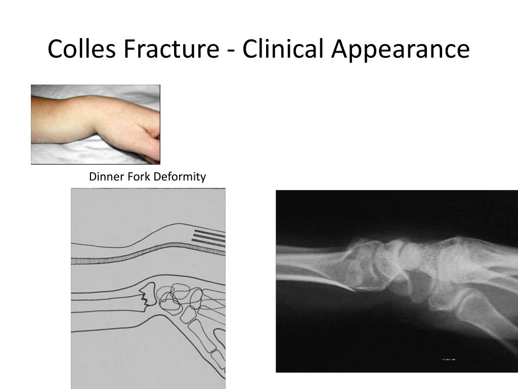
Deformity of the wrist, sometimes called a "dinner fork deformity," which causes it to look crooked and bent. To diagnose a broken wrist, your doctor will give you a thorough physical exam .

DINNER FORK DEFORMITY Mobile Physiotherapy Clinic Ahmedabad Gujarat
On examination, the patient's right wrist is swollen with marked "dinner fork" deformity. There is tenderness over the distal radius and decreased sensation in the distribution of the median nerve. Other Presentations. Fractures of the distal radius can sometimes be associated with open wounds. Irrespective of age, an open wound usually.

Dummies Guide to Colles' fracture Exercise After Cast Removal
The structures distal to the fracture (wrist and hand) are displaced posteriorly. It produces what is known as the 'dinner fork deformity'. Fractures of the radial head - This is characteristically due to falling on an outstretched hand. The radial head is forced into the capitulum of humerus, causing it to fracture.

Food related medical terms Dinner fork deformity
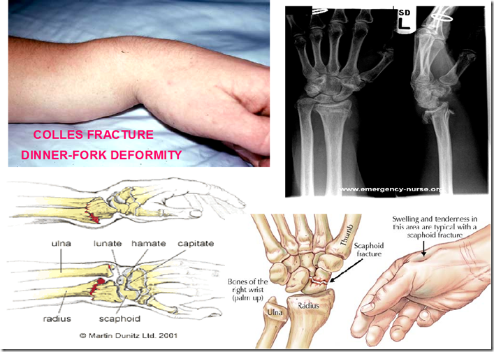
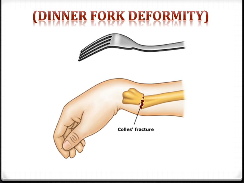
In the distal radius, the term 'Colles' fracture' is still used to describe a fracture in which there is an obvious and typical clinical deformity (commonly referred to as a 'dinner fork deformity') of dorsal translation, dorsal angulation, dorsal comminution (fragmentation) and shortening.
JMSR
Displaced fractures usually present with a 'dinner fork' deformity and require closed reduction and possible surgical fixation. Successfully reduced fractures can be treated non-surgically with immobilisation and radiographic monitoring.

6 Pics Dinner Fork Vs Garden Spade Deformity And Description Alqu Blog
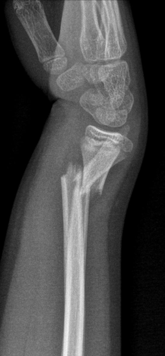
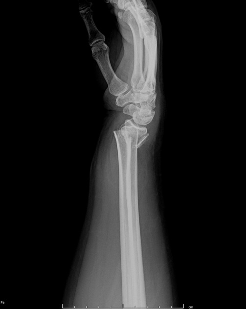
Dinner fork deformity is a term used to describe the appearance of the wrist on a lateral radiograph in cases of distal radial fracture with associated dorsal displacement and impaction, as seen in cases of Colles fracture. The mechanism of injury is usually a fall on an outstretched hand.

PPT Colle’s Fracture PowerPoint Presentation, free download ID1901634
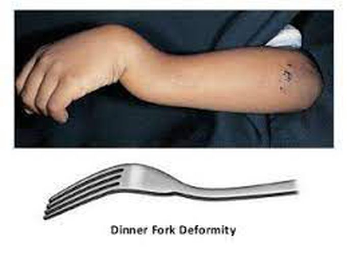
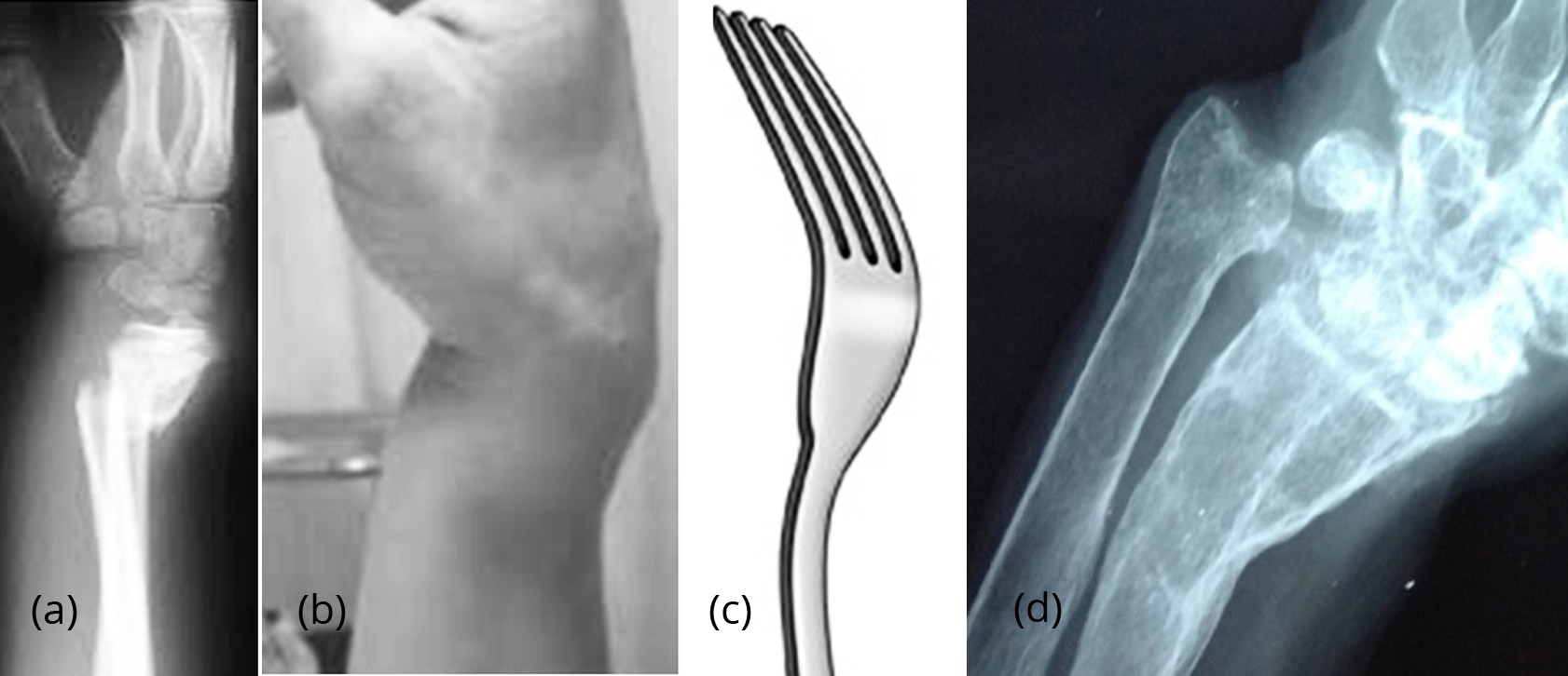
A dinner fork deformity, also known as a bayonet deformity, occurs as the result of a malunited distal radial fracture, usually a Colles fracture. The distal fragment is dorsally angulated, displaced and often also impacted. The term is descriptive, as the lateral view of the wrist is similar to the shape of a fork, seen from the side, tines down.
Colles fracture dinner fork deformity Image
A Colles' fracture is a type of fracture of the distal forearm in which the broken end of the radius is bent backwards. [2] Symptoms may include pain, swelling, deformity, and bruising. [2] Complications may include damage to the median nerve. [1] It typically occurs as a result of a fall on an outstretched hand. [2]

PPT Orthopaedics Tutorial PowerPoint Presentation ID3924770
Displaced fractures usually present with a "dinner fork" deformity and require closed reduction and possible surgical fixation. Successfully reduced fractures can be treated nonsurgically with immobilization and radiographic monitoring.
Dinner fork deformity (wrist) pacs
A Colles Fracture is a complete fracture of the radius bone of the forearm close to the wrist resulting in an upward (posterior) displacement of the radius and obvious deformity. It is commonly called a "broken wrist" in spite of the fact that the distal radius is the location of the fracture, not the carpal bones of the wrist. [1]

Child With Dinner Fork Deformity Annals of Emergency Medicine
The deformity that results from the Colles' fracture is described as a "dinner fork" deformity because of depression at the fracture site, dorsal angulation, and dorsal displacement of the distal radius. History-taking should focus on the mechanism of injury and amount of energy involved.
Dinner fork deformity causes , symptoms & treatment
A dinner fork deformity also identifies as a bayonet deformity, happens as the result of a malunited distal radial fracture, generally a Colles fracture. The distal fragment is dorsally angulated, displaced, and often also impacted. What are the Causes of Dinner fork deformity? Wrist fracture mainly after colle's fracture
PPT Distal Radius Fractures PowerPoint Presentation, free download ID4048273
Objectives: Identify the etiology and epidemiology of Colles fractures. Outline the appropriate history, physical, and evaluation of a patient with a Colles fracture. Review the treatment and management options available for Colles fractures.
Classical Dinner Fork Deformity of Colles' Fracture of Wrist
The clinical presentation of Colles fracture is frequently described as a dinner fork deformity - distal fracture of the radius causes posterior displacement of the distal fragment, causing the forearm to be angled posteriorly just proximal to the wrist.

Figure 1 from The Dinner Fork Deformity Semantic Scholar
Overview A Colles' wrist fracture occurs when the radius bone in your forearm breaks. It's also known as a distal radius fracture, transverse wrist fracture, or a dinner-fork deformity of.